Bungaro-toxin marker for histology:
A powerful tool for the quantification and evaluation of the Acetyl-Choline receptors on the NMJ.
Dr. Emmanuel Loeb
Introduction:
The neuromuscular junction (NMJ) involves a complex structure whose function is to convey action potentials from the motorneuron (MN) to skeletal muscle, resulting in stimulated contraction of the muscle.
Bungarotoxin (BTX-488), the toxin that specifically binds to acetylcholine receptors (AchRs) on the surface of muscle fibers can show the receptor morphology for histological evaluation.
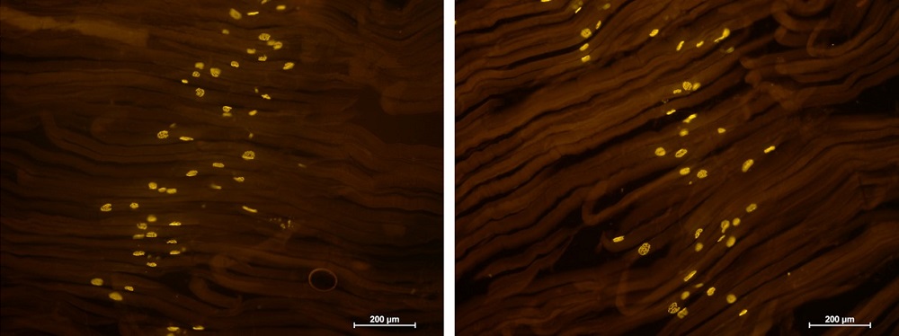
An example of Bungarotoxin receptors stain in a myasthenia gravis study of a mouse model.
Tissue preparation protocol for Acetylcholine analysis
Work protocol:
Organ/Tissue Collection and Fixation
- Samples of striated muscles (gastrocnemius and diaphragm) can be harvested from mice in different disease models.
Bungarotoxin staining for Acetylcholine receptors (AChRs) labelling
- Following isolation of the gastrocnemius and the diaphragm, the whole muscles are incubated for 20 mins with tetramethlrhodamine conjugated -bungarotoxin (BTx, 1:100, Invitrogen) to label AChRs.
- Muscles are washed briefly with PBS and fixed with 2.5% PFA for additional 20 min and then were cryoprotected in 30% sucrose PBS solution for ON. The next day, muscles are placed in molds with OCT and were frozen and kept at -20°C until further used.
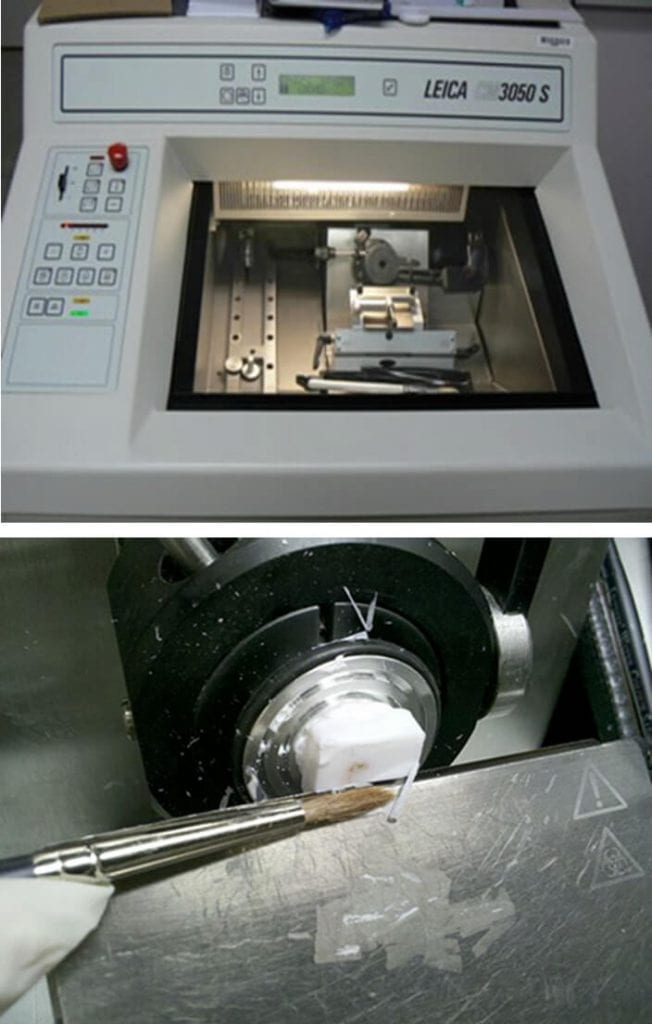
Slide Preparation
- 30 μm thick frozen sections are cut by cryostat in a plane parallel to the surface of the muscle and mounted on super-frost+ slides (20 sections per muscle). Sections are dried and covered, using aqua-mount mounting solution.
Microscopic examination and photography
- Stained sections are examined by fluorescent microscope equipped with Plan Apo objectives connected to a CCD camera. Digital images of the AChRs, 6-10 fields of 0.2 mm2 from each muscle are collected. Analysis is performed in X6 and X12 magnifications.
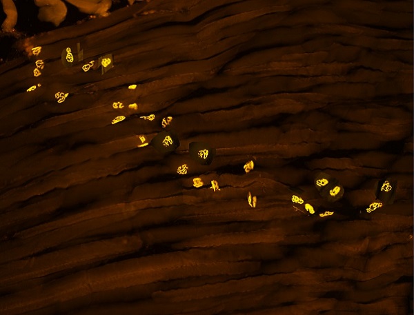
The analysis process of Acetyl-Choline receptors
Morphometric analysis of the immunofluorescence (IF) reaction for labelled AChRs
Morphometry of the area of the AChRs and the percent of the labelled area can be performed, using image analysis tools: The number and area of the receptors are analyzed using image pro v10 and the MATLAB software. The area of the labeled receptors is held as a parameter for the number of receptor sites per area of 0.2 mm2. The percentage of intact receptors per area of 0.2 mm2 is held to quantify the grade of the intact morphology of the individual receptors per area unit.
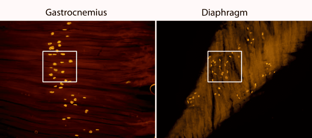
Area of 0.2 mm2 marked in a white square per muscle, from each sample 6-8 such areas were calculated for the number of receptors and the % of the receptors size (morphology).
Histological examples of stained sections:
Bungarotoxin stain in gastrocnemius muscle of healthy animal, volume and morphology are perfectly intact

Bungarotoxin stain in gastrocnemius muscle of affected animal, irregular number and morphology.