Digital morphometry quantification of dopamine activity in a 6-hydroxydopamine (6-OHDA) Parkinson model in mice
Loeb E¹, Zuckerman A¹, Margalit R², Orgad U³. ¹Patho-Logica, Golda Meir Str 7, Scientific Park, Ness-Ziona, Israel. ²Science In Action, Sapir Str. 5, Scientific park, Ness-Ziona, Israel. ³Hebrew University, Koret School for veterinary medicine, Rehovot, Israel
Introduction:
- Parkinson’s disease is a progressive neurodegenerative disorder, that is primarily characterized by degeneration of the dopaminergic neurons of the nigrostriatal pathway.
- The neurotoxin 6-hydroxydopamine (6-OHDA) continues to constitute a valuable tool to model Parkinson’s disease in rats and mice (1).
Aim:
- To quantify the amount of damaged dopamine producing cells and fibers by digital morphometric tools, compared to scale traditional semi-quantitative analysis based on score scale (2).
Materials and methods:
- Intrastriatal injection of 6-OHDA (3 μl) was applied into the right hemisphere of mice (n=5), at the following coordinates: (i) AP=+1.0, L=−2.1, DV=−2.9;(3). The left hemisphere served as control.
- Animals were sacrificed 8 weeks later. Brains were dissected to obtain sections from the striatum (ST) and the Substantia Nigra Pars Compacta (SNC).
- Five mµ thick paraffin sections were cut and stained for tyrosine hydroxylase (TH) activity, a marker for dopamine production, using immunofluorescence histochemistry.
- The intensity of the reaction was measured by computerized morphometry (Image Pro Plus 6.3), and compared to the semi-quantitative analysis, (score scale).
Semi-quantitative analysis (score scale):
– = Normal TH expression
+ = Mild TH reduction
++ = Moderate reduction
+++ = Severe TH reduction
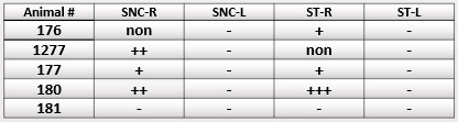
Table 1: Semi quantitative analysis of TH activity based on a score scale. Note the reduction of TH in the treated side (R) compared with the untreated hemisphere (L).
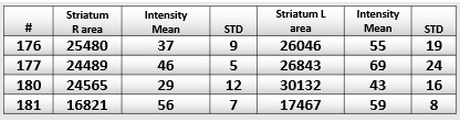
Table 2: Quantitative analysis of TH activity using digital morphometry. Note the decrease in the treated side (R) compared with the untreated hemisphere (L).
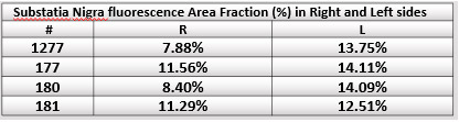
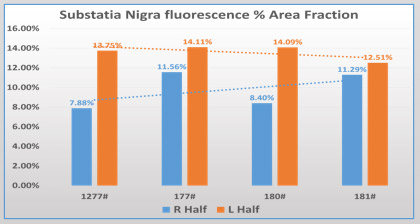
Table 3: Histogram of % of TH positive cells in SNC
Results:
- Morphometric analysis: The quantitative analysis showed a pronounced decrease in the intensity of the reaction in the ST in the right hemisphere, compared to the left un-treated control.
- In the SNC, the activity of TH in the right hemisphere was lower compared to the control left hemisphere.
- Semi quantitative analysis: A decrease of TH in the ST and SNC was observed, compared to the left hemisphere.
- The morphometric analysis could detect slight changes that were not shown by the semi-quantitative analysis (for example see animal 181).
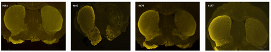
Figure 1. Coronal brain sections at the level of the striatum, showing the activity of TH expressed by fluorescence. Note the reduction in TH expression in the right hemisphere compared to the left control. The digital Intensity analysis is presented in the histogram below.
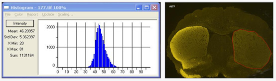
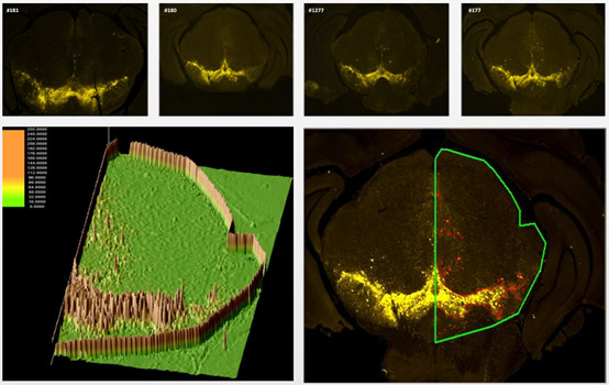
Figure 2. Coronal brain sections at the level of the substantia nigra, showing the intensity of TH activity. Note the reduction of TH expression (fluorescence) in the right hemisphere, compared to the left side. The morphometric digital surface analysis of TH positive cells is presented in the left image.
Summary and Conclusions:
- A clear damage in the striatum and in the substatia nigra was shown following injection of 6-OHDA , expressed by decrease in TH activity.
- An accurate number for the TH activity was obtained, by using a digital morphometric tool, in contrast to the semi-quantitative method.
- The digital quantification of histological markers is highly accurate compared to the traditional semi-quantitative method and therefore recommended especially for efficacy studies, where slight differences between treatments are essential.
References:
- Schober A. Classic toxin-induced animal models of Parkinson’s disease: 6-OHDA and MPTP, Cell & Tissue Res 2004; 318: 215–224.
- Jansen JA et al. Semi-quantitative and qualitative histologic analysis method for the evaluation of implant biocompatibility. J Invest Surg; 1994,;7:123-34.
- Fischer K et al. In vivo quantification of dopamine transporters in mice with unilateral 6-OHDA lesions using [11C]methylphenidate and PET. Neuroimage 2012; 59:2413-22.
- Mori S et al. Lack of interleukin-13 receptor α1 delays the loss of dopaminergic neurons during chronic stress. J Neuroinflammation 2017: 14:88.