Comparison of methods to evaluate cuprizone-induced demyelination in mice
Litinetsky La; Belinson, Ha; Geva, Ma; Blumenfeld – Katzir, Tb; Loeb, Ec; Amit- Romach, Ea; Orbach, Aa. aDiscovery & Product Development, Global Research & Development, Teva Pharmaceutical Industries Ltd., Netanya, Israel; b ImagingQ, Ramat Gan, Israel. c Patho – Logica Ltd., Golda Meir St 7 ,Scientific Park, Ness Ziona, Israel.
Support for this study was provided by Teva Pharmaceuticals, Israel.
INTRODUCTION
The Cuprizone (CPZ) mouse model is a well-established method for toxic demyelination that has been used in several magnetic resonance imaging (MRI) studies. In this model, mice are fed with CPZ, leading to oligodendrocyte death and subsequent demyelination, most profoundly observed in the corpus callosum (CC). The loss of white matter in the CC can be quantified either by histological tools or imaged by MRI.
MRI analysis is a powerful non-invasive method to evaluate the myelination state of the nervous system and, hence, is highly useful to track the progression of the model and any protective treatment.
The aim of the study was to evaluate the capability of a compact high-performance MRI system (1Tesla aspectimaging®) to assess demyelination of the CC in the cuprizone model.
METHODS
Animals and treatments: Thirty 8-12 week old male C57BL/6 mice were fed for 6 weeks with either normal chow or a diet containing 0.3% cuprizone mixed into pelleted rodent chow (1,2). To evaluate if treatment related changes can be detected, we treated mice with test compound X (TCX) which shows beneficial effect in the model and can serve as a positive control. TCX was dosed at 25 mg/kg PO QD. Following 6 weeks of treatment, mice were scarified and perfused in PFA. Brain in suckle were kept prepared for MRI analysis. Thereafter, brains were excised from the skull and prepared for histology. All experimental procedures conformed to accepted ethical standards for use of animals in research and were in accordance with the Committee for the Care and Use of Experimental Animals guidelines and approved by the Teva Institutional Animal Care and Use Committee.
MRI: Ex vivo brain samples were scanned using a 7 Tesla MRI scans (Bruker Biospec 7T/30 Scanner, SCAN@TAU Center) and 1 Tesla (M3TM compact low MRI machine, aspectimaging®) according to the following protocols:
7 Tesla 1 Tesla
Resolution: 100 micron Resolution: 100 micron
No. of slices: 12 No. of slices: 9
Slice thickness: 0.6 mm Slice thickness: 0.9 mm
Time scan: 26 min Time scan: 120 min
Assessment of the demyelination of the CC of mice was performed by volumetric analysis of the CC (3)
Histopathology: Brains were trimmed in a standard position for all the samples, using the chiasma opticum as a reference landmark. The frontal half brain was processed for paraffin embedding for immunofluorescent staining of MBP and the rest remained in the fixative until cut in a cryostat for Black Gold (BG) staining for myelin.(4) Stained sections were examined and photographed by using Plan Fluor objectives (40x) connected to a camera (DS-Qi1, Nikon). Digital images were collected and analyzed using Image-Pro software.
* All samples were analyzed by these methods in a blind manner.
RESULTS
- MRI analysis of loss of CC volume in the CPZ model
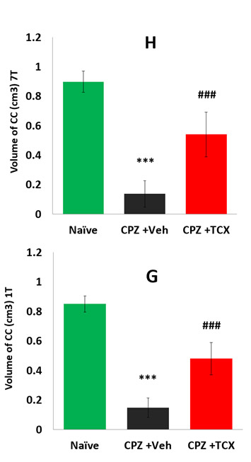
Using specific protocols for the 7 Tesla we found that brains from CPZ- treated mice showed a pronounced decrease in CC volume, compared to naive brains (Fig. 1A, B, G). In brains from CPZ-fed mice that were treated with TCX, a drug that ameliorates CPZ-induced demyelination, an intermediate grade of demyelination and CC volume loss was exhibited (Fig. 1C, G). In addition, using unique protocols for the 1 Tesla we were able to measure similar loses in the volume of the CC to those obtained with the 7 Tesla (Fig. 1).
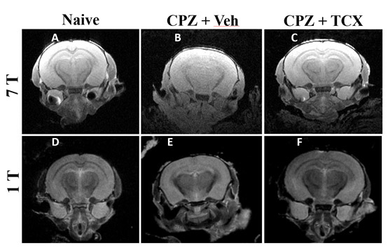
Figure 1: CC volumetric loss is similar in 7 Tesla and 1 Tesla MRI
***p<0.001 – as compared to Naïve group;
### p<0.001 – as compared to CPZ+ Veh, by ANOVA, Fisher’s LSD
- Histopathology analysis of loss of fibers in the CC in the CPZ model
To further assess the brain pathology we performed MBP immunofluorescent and Black Gold (BG) histological staining. Using BG staining we found that the CC from cuprizone treated mice showed a pronounced decrease in the positive fiber staining, compared to naive brains (Fig. 2A, B, H). In brains from CPZ-fed mice that were treated with TCX, the loss of fibers was significantly ameliorated (Fig. 2C, H). In addition, using MBP immunofluorescent similar results were obtained, by which CPZ treatment decreased MBP positive fibers and TCX treatment ameliorated this effect (Fig. 2G). Important to note, the histological analysis revealed comparable results to those obtained by the MRI analysis of the CC volume loss.
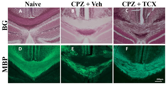
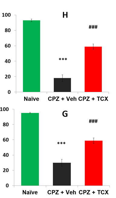
Figure 2: BG and MBP fiber loss analysis in the CPZ model
***p<0.001 – as compared to Naïve group ;
### p<0.001 – as compared to CPZ+ Veh, by ANOVA, Fisher’s LSD
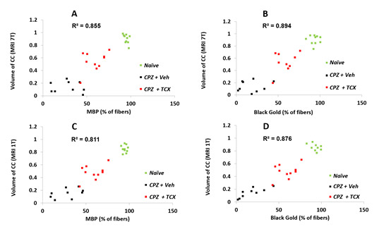
III. Correlation of MRI and histopathology analysis in the CPZ model
Analysis of the correlation of the MRI and histological analysis revealed that, both MRI 7 Tesla and 1 Tesla correlate similarly with the histological data. Important to note, BG correlation with the MRI analysis seemed to have higher correlation but it was not significantly different.
CONCLUSIONS
The results of the present study show that both MRI methodologies and the histopathology provide comparable readouts for the grade of demyelination of the CC in the cuprizone mouse model. Moreover, the MRI methodologies and the histopathology analysis show significant correlation. Thus, we believe that any of these methodologies could serve as a screen of efficacy in these models. Moreover the same sample could be used for the initial MRI screen and in depth histopathological evaluation.
REFERENCES
- Torkildsen U, Brunborg LA, Myhr K-M, BL. The cuprizone model for demyelination. ActaNeurol Scand 2008: 117 (Suppl. 188): 72-76.
- Skripuletz et al. Cortical Demyelination Is Prominent in the Murine Cuprizone Model and Is Strain-Dependent. Am J Pathol 2008, 172 (4): 1053-106.
- Matsushima, Glenn K., and Pierre Morell. The neurotoxicant, Cuprizone, as a model to study demyelination and remyelination in the central nervous system. Brain Pathology 2001(11.1) : 107-116.
- Franco-Pons, Neus, et al. Behavioral deficits in the Cuprizone-induced murine model of demyelination/remyelination. Toxicology Letters 2007 (169.3): 205-21.